Designing a new diagnostic system for the most common cancer among Iranian women by scanning with a high-frequency electromagnetic wave probe.
According to the public relations report of University of Tehran, the results of this research, which was published as High-Frequency (30 MHz-6 GHz) Breast Tissue Characterization Stabilized by Suction Force for Intraoperative Tumor Margin Assessment in Diagnostics magazine, show that with the help of electromagnetic waves with high frequency High (30 MHz to 6 GHz) can detect breast cancer.
Dr. Mohammad Abdulahad, a faculty member of the School of Electrical and Computer Engineering, University of Tehran and head of the Electronic Cancer Research Center of University of Tehran and Tehran University of Medical Sciences(TUMS), said: We are completing the cycle of diagnosing breast cancer masses and margins before, during and after surgery and We are an operating room and a pathology room, and this system is a ring of this cycle.
He, who is the inventor of the CDP cancer diagnostic probe (which is now an approved intraoperative diagnostic device and is being used in clinical centers) and the winner of the Mustafa Award in 2019, has started organizing research to diagnose the most common cancer among Iranian women. He announced the help of high frequency electromagnetic waves in the form of two master's theses and added: by researching and using the method of electrical stimulation at GHz frequencies and calculating and measuring the scattering parameters, an attempt is made to use the freshly removed tissue from the patient's body without any preparation process. , additional information about the location of tissue suspected of cancer to be given to the pathologist in a short period of time in order to reduce the time and cost of diagnosis in the pathology laboratory.
Dr. Abdulahad stated: Specifically, the purpose of this dissertation is the electrical characterization of cancerous and healthy breast tissues at frequencies of 30 MHz to 6 GHz, or the so-called biology of the gamma dispersion region.
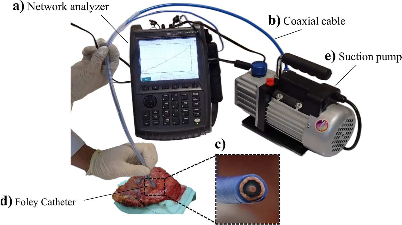
A faculty member of the School of Electrical and Computer Engineering of University of Tehran said about the measurement and diagnosis method: One of the basic differences between cancer and healthy cells is the amount of water molecules inside the cell connected to the proteins of the inner part of the membrane. This difference, which causes the dipole resonance of water molecules, is different between healthy and cancer cells. The measurement method in this research is that with the help of network analyzer and coaxial probe, the dispersion parameter S11, which is the ratio of return power to transmitted power, was measured for breast tissue. One of the most important factors that can greatly increase the measurement error is the way the probe is attached to the tissue. In order to solve this problem, a vacuum pump device was used. The measurement method can be seen in Figure 1.

Figure 1: How to measure using this method and introducing its different parts
Saying that this project was defined in three phases, the associate professor of the Faculty of Electrical and Computer Engineering of University of Tehran added: In the first phase, this stimulation was applied to healthy and cancerous mouse tissue, and after the investigations, the difference between the scattering parameters for healthy and cancerous mouse tissue It was visible. which gave us the confidence to enter the next phases.
He said: The second phase was to check the correct detection rate of this device for healthy and cancerous breast tissue and to determine the demarcation for all types of breast tissue. For this purpose, measurements were made on small breast tissues and by determining different boundaries and numerous statistical analyses, finally by determining the boundary of -7.25 dB for healthy and cancerous breast tissues, the accuracy of this device for detecting healthy and cancerous breast tissue was 87 It was % percent. In this phase, a total of 127 samples from 19 patients were examined.
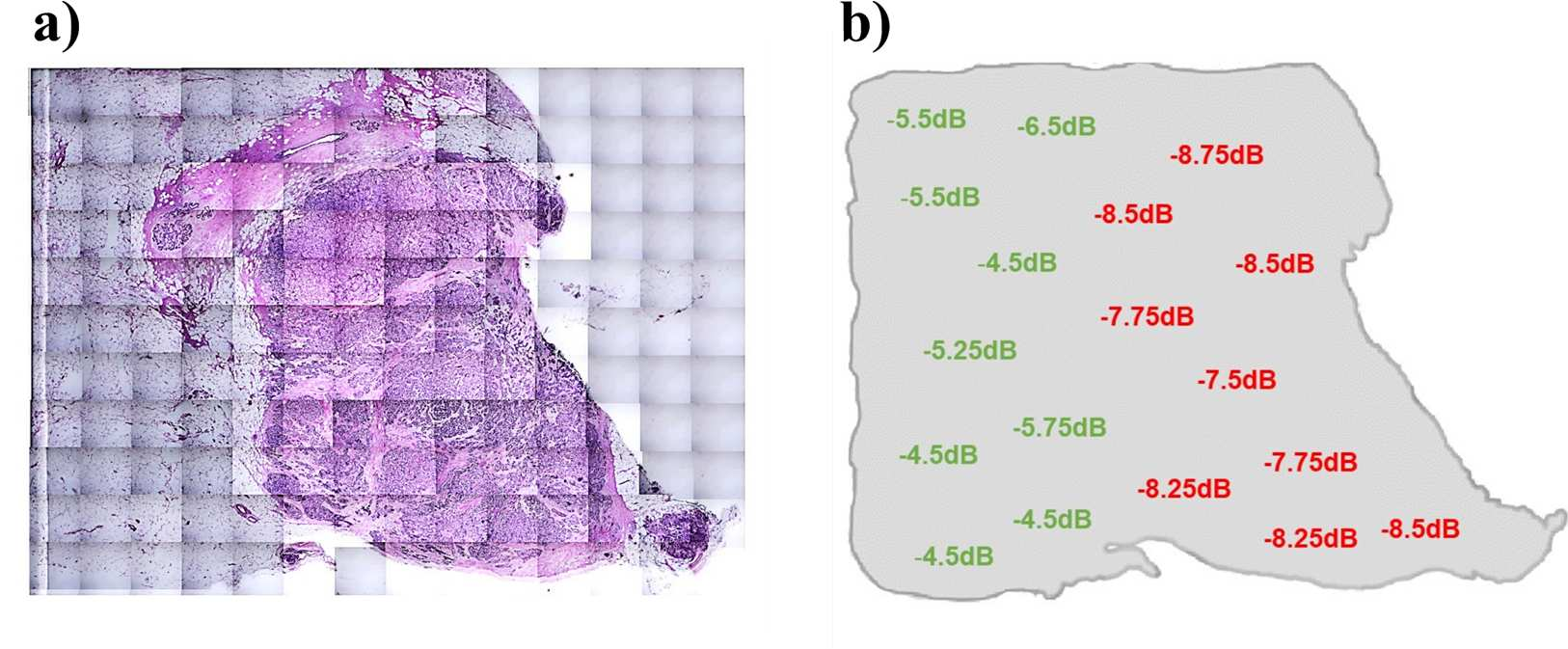
In explaining the final phase of this project, the affiliated faculty member of Tehran University of Medical Sciences added: In the final phase, the most important goal of this project was to scan the breast tissue surface without any invasive tools. In this phase, after ensuring the correctness of the method and design and determining the exact demarcation of healthy and cancerous breast tissue, we went to the complete examination of the tumor margin. This phase was defined to apply and determine the healthy and cancerous margins of tumors, which is one of the biggest concerns of surgeons, in which instead of measuring from only one point and taking small samples, this time the tissues of the tumor margins with dimensions of approximately 3 x 2 cm square was selected and an average of 20 measurements were performed from each sample. The exact location of each measurement was marked with special pathology ink so that the results of the measurement and the pathology were matched. In this phase, 86 samples were collected from 22 patients. The results can be seen in Figure 2.

Figure 2: Comparison of surface scan results using this method and H&E slides
Dr. Abdulahad noted: To summarize, we need to examine the theory of this phenomenon. In most of the articles reviewed in this treatise, the difference in the amount of water in the cells and between the tissues was considered to be the reason for the difference in the response of the dielectric characteristics of healthy and cancerous cells.
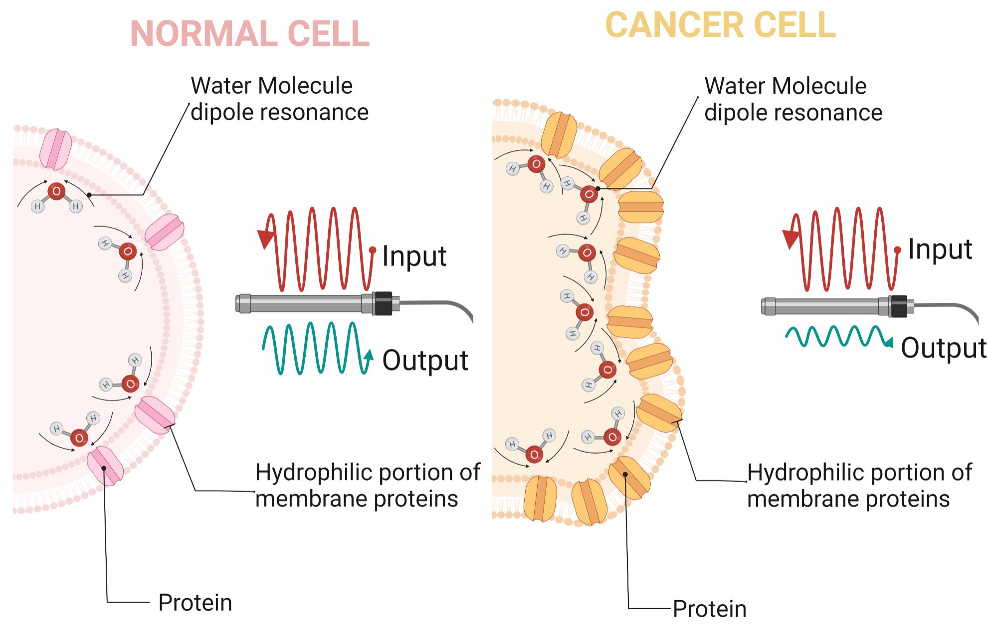
He said: The rapid growth and proliferation of cancer cells leads to the increase and expansion of proteins (including membrane proteins) compared to healthy cells. The mentioned proteins on the surface of the cell membrane molecules absorb more water as a dominant component in the cell environment to form bound-water. These dipoles can be oriented by an oscillating electric field at frequencies below 20 GHz. In this way, the higher amount of bound-water increases the absorption of electromagnetic energy and cellular response in frequencies. Figure 3 shows a schematic of the difference between the structure of healthy and cancerous cells.

Figure 3: Schematic of the difference between the structure of healthy and cancerous cells and the response to electromagnetic wave at high frequency
This research is the result of the master's thesis work of Hadi Mokhtari Dulatabad, a student of electrical engineering.
Interested parties and researchers can refer to the link https://doi.org/10.3390/diagnostics13020179 to get the original article.

Your Comment :